41 electron micrograph labeled
Quintuple labeling in the electron microscope with genetically ... by D Cruz-Lopez · 2018 · Cited by 8 — In electron microscopy, however, true genetic encoded multilabeling ... (c) Electron micrograph of the same HRP-labeled dendrite as in (b). plant cell label electron micrograph Diagram | Quizlet plant cell label electron micrograph Diagram | Quizlet plant cell label electron micrograph 3.3 (3 reviews) + − Learn Test Match Created by June_bee743 Terms in this set (7) chloroplast ... cell wall ... plasma membrane ... golgi apparatus ... nucleus ... vacuole ... mitochondria ...
What is Electron Microscopy? - UMASS Medical School Electron microscopy (EM) is a technique for obtaining high resolution images of biological and non-biological specimens. It is used in biomedical research to investigate the detailed structure of tissues, cells, organelles and macromolecular complexes. The high resolution of EM images results from the use of electrons (which have very short ...
Electron micrograph labeled
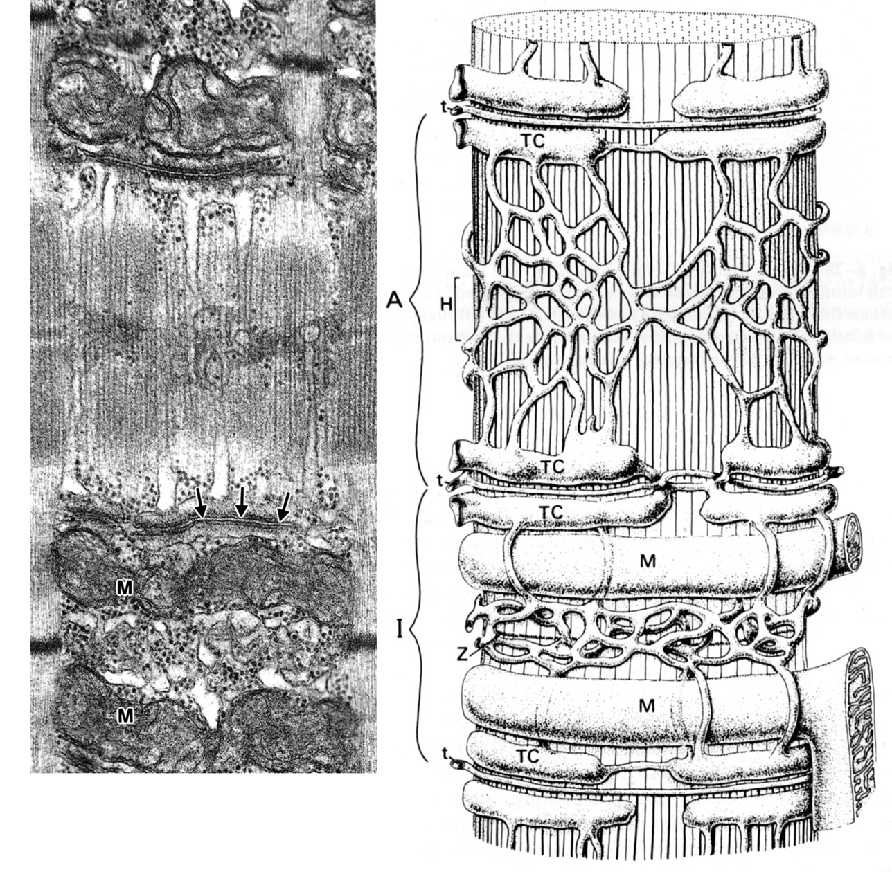
Skeletal Muscle EM - Yale University Skeletal Muscle EM This is an electron micrograph of a longitudinal section of skeletal muscle. First, focus on the components in the different bands. The I-Band contains actin filaments and is bisected by the Z-disk. The A-Band contains myosin and actin filaments. The M-line is a disc-like zone where myosin filaments are crosslinked. Electron Micrographs - University of Oklahoma Health Sciences Center Electron Micrographs Below is a collection of electron micrographs with labelled subcellular structures that you should be able to identify. Also, be sure to observe any electron micrographs which are made available in the laboratory by the instructor. Transmission electron microscopy DNA sequencing - Wikipedia In order for DNA to be clearly visualized under an electron microscope, it must be labeled with heavy atoms. In addition, specialized imaging techniques and aberration corrected optics are beneficial for obtaining the resolution required to image the labeled DNA molecule.
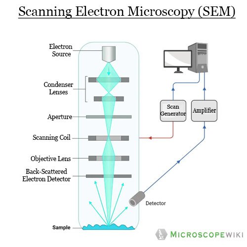
Electron micrograph labeled. Electron Microscope- Definition, Principle, Types, Uses, Labeled Diagram An electron microscope is a microscope that uses a beam of accelerated electrons as a source of illumination. It is a special type of microscope having a high resolution of images, able to magnify objects in nanometres, which are formed by controlled use of electrons in a vacuum captured on a phosphorescent screen. Transmission Electron Microscope (With Diagram) - Biology Discussion Transmission Electron Microscope (With Diagram) In this article we will discuss about the design of transmission electron microscope, explained with the help of a diagram. In TEM a finely focused beam of electrons from an electron gun is passed through a specially prepared ultra thin section of the specimen. The beam is focused on a small area ... Sperm Under Microscope with Labeled Diagram - AnatomyLearner The normal light microscope easily shows these stereocilia of the epididymal ducts. But, the electron microscope will show a clear view of these stereocilia. You will also see the agranular endoplasmic reticulum, lysosome, and prominent Golgi bodies in these lining epithelia of the epididymis (with an electron microscope). Scanning Electron Microscope (SEM)- Definition, Principle, Parts ... The first Scanning Electron Microscope was initially made by Mafred von Ardenne in 1937 with an aim to surpass the transmission electron Microscope. He used high-resolution power to scan a small raster using a beam of electrons that were focused on the raster.
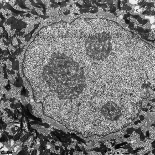
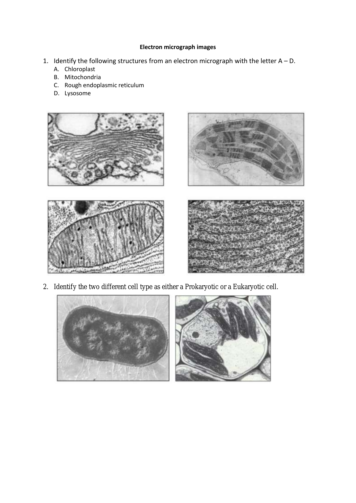
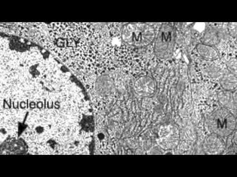
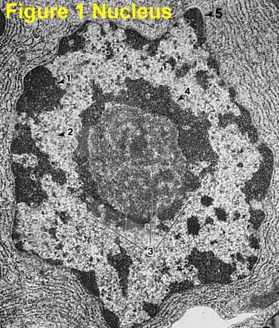
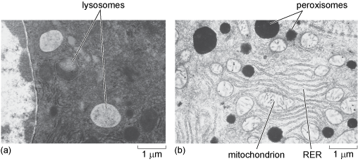
Electron Micrographs of Cell Organelles | Zoology - Biology Discussion The Electron Micrograph of Nucleus: This is an electron micrograph of nucleus. (Fig. 17 & 18): (1) Nucleus was discovered by Brown (1831). (2) It is a characteristic entity of almost all eukaryotic cells except mammalian RBCs. (3) The nucleus is generally one but may also be two, four or many. Home.php Lab - Yale University Given an electron micrograph of a skeletal or cardiac muscle cell, students should be able to label the components of the sarcomere; Introduction. Muscles are multicellular contractile units. They are divided into three types: ... Smooth Muscle Cell Electron Micrograph. The thick (myosin) and thin (actin) filaments are scattered throughout the ... Electron Microscope Images That Show The Power of Electron Microscopes An electron microscope is a sophisticated and versatile imaging device that can be used to study live and fixed organic and inorganic materials, as well as observe their transformation over a certain period of time. Electron microscopes offer us the capability to study things right down to the atomic level. Microscope Types (with labeled diagrams) and Functions Electron microscope labeled diagram The different types of electron microscopes are: Transmission Electron Microscope Scanning Electron Microscope Reflection Electron Microscope Scanning transmission electron microscope Scanning tunneling microscopy Electron microscope functions: Semiconductors and Data Storage Industry Failure Analysis
Electron microscope - Wikipedia An electron microscope is a microscope that uses a beam of accelerated electrons as a source of illumination. As the wavelength of an electron can be up to 100,000 times shorter than that of visible light photons, electron microscopes have a higher resolving power than light microscopes and can reveal the structure of smaller objects. A scanning transmission electron microscope has achieved ... Labeling the Cell Flashcards | Quizlet Label the transmission electron micrograph of the nucleus. membrane bound organelles golgi apparatus, mitochondrion, lysosome, peroxisome, rough endoplasmic reticulum nonmembrane bound organelles ribosomes, centrosome, proteasomes cytoskeleton includes microfilaments, intermediate filaments, microtubules Identify the highlighted structures Histology and Microscope Slide Labels & Tape | EMS Electron Microscopy Sciences Categories. General Supplies for Specimen Preparation Bottles and Beakers ... Description: Microscope End Label SES P-16-RC Pathology: Pack: 1000 Roll: Price: $34.00: Add to Quote: Add: Cat #: 77020-05: Description: SLIDE LABEL, STANDARD ROLL: Pack: 1000 Roll: Electron Microscope Principle, Uses, Types and Images (Labeled Diagram ... Electron Microscope Principle, Uses, Types and Images (Labeled Diagram), Price Electron Microscope By Editorial Board October 3, 2022 The advances in technology have enabled the development of powerful microscopes to view the samples at a nanometer level and thus were born the electron microscopes.
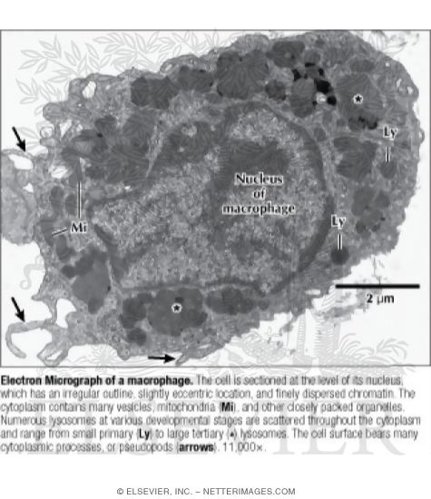
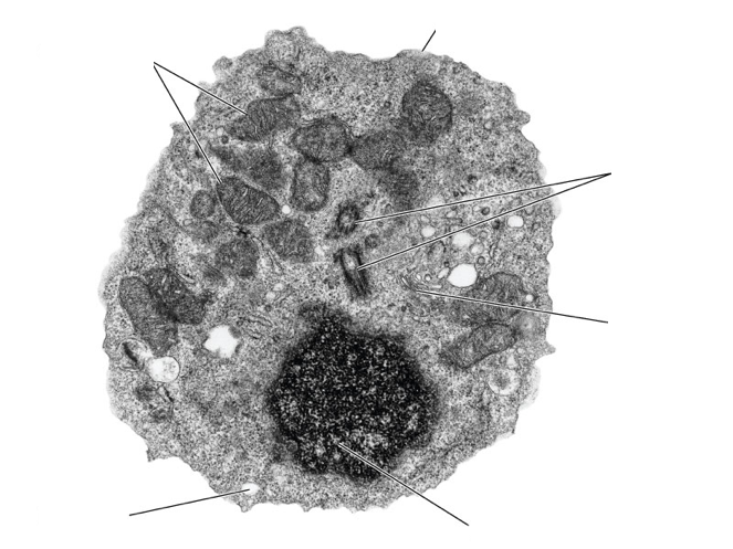
Electron Micrograph of a Macrophage
Clarifying electron micrograph labeling - JAMA Network by C Marwick · 1986 — Two years ago, a JAMA news article on acquired immunodeficiency syndrome (AIDS) was accompanied by three electron micrographs labeled ...
Biology 130 Lab 2 - Electron Micrographs
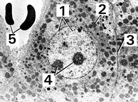
Virtual EM Micrograph List | histology - University of Michigan 021. Plasma Cell: This electron micrograph shows a typical secretory cell, a plasma cell, which secretes immunoglobulin protein. Many of the major types of cellular organelles are visible in this image. In the nucleus, areas of euchromatin and heterochromatin can easily be identified. Virtual Slide.
Electron Micrograph of Actin and Intermediate Filaments In ...
Electron micrograph - definition of electron micrograph by The Free ... electron micrograph n (Biology) a photograph or image of a specimen taken using an electron microscope Collins English Dictionary - Complete and Unabridged, 12th Edition 2014 © HarperCollins Publishers 1991, 1994, 1998, 2000, 2003, 2006, 2007, 2009, 2011, 2014 Want to thank TFD for its existence?
The Cell: The Histology Guide
Electron microscopes - Cell structure - Edexcel - BBC Bitesize the transmission electron microscope (TEM) is used to examine thin slices or sections of cells or tissues the scanning electron microscope (SEM) has a large depth of field so can be used to...
Histology Laboratory Manual
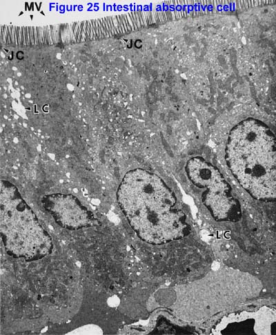
PDF Electron Micrographs (EMs) for laboratories in A215, Basic Human ... - IU of the micrograph. There are distinct differences between cilia and microvilli to be seen in electron micrographs: - Cilia are larger (the cilium labeled C about 2.5 microns along its length is probably 5 to 10 microns long); - Cilia contain microtubules, by which they can move. These are seen in longitudinal sections of cilia as dark

1. On the following Transmission Electron Micrograph of a ...
What Is an Electron Microscope (EM) and How Does It Work? - VHA ... Today there are two major types of electron microscopes used in clinical and biomedical research settings: the transmission electron microscope (TEM) and the scanning electron microscope (SEM); sometimes the TEM and SEM are combined in one instrument, the scanning transmission electron microscope (STEM):
A tour of the cell: View as single page
Neuron under Microscope with Labeled Diagram - AnatomyLearner Neuron under Microscope with Labeled Diagram. 03/11/2022 31/03/2022 by anatomylearner. The structural and functional unit of the nervous system is the neuron that may easily observe under a light microscope. Neurons may vary considerably in size, shape, and other features. ... So, when you are observing the neuron under the electron microscope ...

Electron micrograph of section of a portion of rat glomerulus ...
Labels, Electron Microscopy Sciences | VWR Labels, Electron Microscopy Sciences; ... Limit label waste by printing only the quantity required. Labels are now oriented vertically and are compatible with the new LabelWriter 450. Tubes labeled with Direct Thermal CryoTags easily slide in and out of centrifuge rotors without binding. Users of Direct Thermal Tough-Tags will be happy to know ...
7,415 Electron Micrograph Stock Photos, Pictures & Royalty ...
Electron Microscope-Definition, Principle, Types, Uses, Labeled Diagram An electron microscope uses an accelerated electron beam to illuminate and is called an electron microscope. It is a particular style of microscope with very sharp pictures that can magnify things down to the nanometer scale. Electrons are precisely guided to form the pictures in a vacuum, which are then captured on a phosphorescent screen.
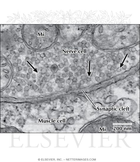
Electron Micrograph of Synaptic Vesicles at a Neuromuscular ...
Electron Micrograph of a Lymphocyte - netterimages.com Electron Micrograph of a Lymphocyte. Variant Image ID: 12970. Add to Lightbox. Email this page. Link this page. Print. Please describe! how you will use this image and then you will be able to add this image to your shopping basket.
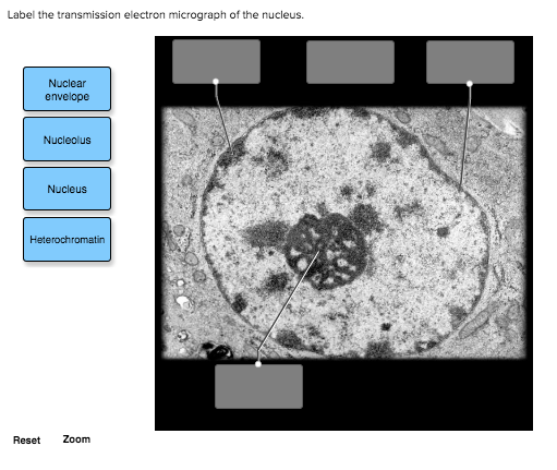
Solved Label the transmission electron micrograph of the ...
Solved QUESTION 32 The figure below shows an electron - Chegg Expert Answer ANSWER (32). Point B will move … View the full answer Transcribed image text: QUESTION 32 The figure below shows an electron micrograph of a skeletal muscle fiber, where various points, and regions, along the sarcomare have been labeled. Which of the following statements is True regarding muscle contraction?
The Biological bulletin. Biology; Zoology; Biology; Marine ...
Label This Transmission Electron Micrograph Of A Relaxed ... - Blogger Electron micrographs of relaxed and contracted muscle fibres. Provide the labels for the electron micrograph in figure 18.5. (b) section through a muscle in the extended condition (140 % of whole muscle resting length). Label the following image using the terms provided.
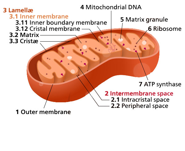
1.7 Mitochondria – Plant Anatomy and Physiology
Transmission electron microscopy DNA sequencing - Wikipedia In order for DNA to be clearly visualized under an electron microscope, it must be labeled with heavy atoms. In addition, specialized imaging techniques and aberration corrected optics are beneficial for obtaining the resolution required to image the labeled DNA molecule.
cell and organelles Dr.Jastrow's electron microscopic atlas
Electron Micrographs - University of Oklahoma Health Sciences Center Electron Micrographs Below is a collection of electron micrographs with labelled subcellular structures that you should be able to identify. Also, be sure to observe any electron micrographs which are made available in the laboratory by the instructor.
Electron Micrographs
Skeletal Muscle EM - Yale University Skeletal Muscle EM This is an electron micrograph of a longitudinal section of skeletal muscle. First, focus on the components in the different bands. The I-Band contains actin filaments and is bisected by the Z-disk. The A-Band contains myosin and actin filaments. The M-line is a disc-like zone where myosin filaments are crosslinked.
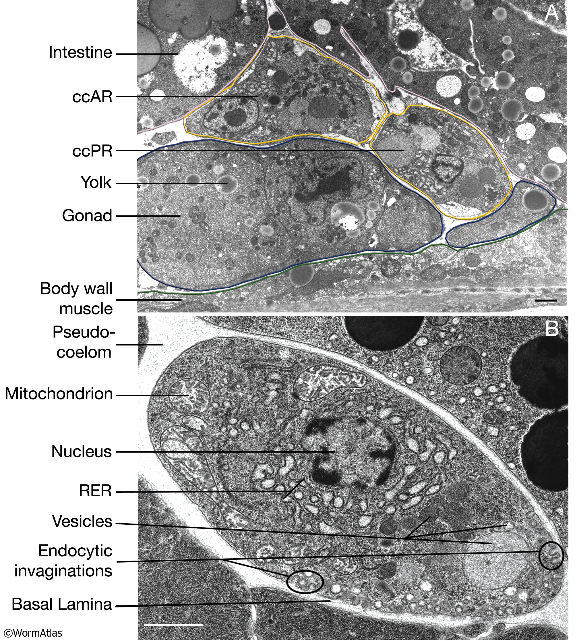
CcFIG 5 Legend
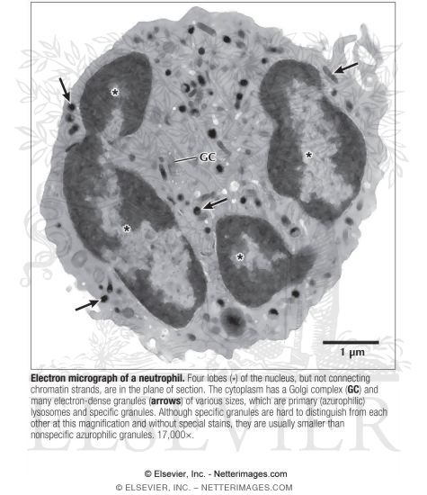
Electron Micrograph of a Neutrophil

DP Biology: Ultrastructure of cells quiz 1.2

Nonmalignant Disorders of Leukocytes Part 1
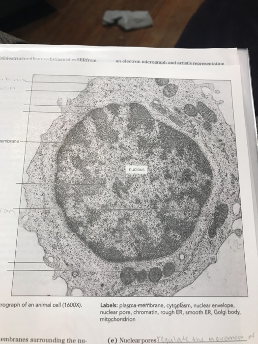
Solved label the ectron micrograph of an animal cell. | Chegg.com
Topic 1.2 Ultra-Structure of Cells - AMAZING WORLD OF SCIENCE ...
Electron Microscope Principle, Uses, Types and Images ...
Electron micrograph
animal cell electron micrograph labelling Diagram | Quizlet

7,415 Electron Micrograph Stock Photos, Pictures & Royalty ...
2.3 Eukaryotic cell - BIOLOGY4IBDP
2.3.3 Identify structures from electron micrographs of liver cells
A tour of the cell: View as single page

Cell Micrographs | BioNinja
Electron Micrographs

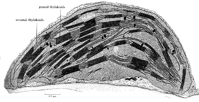
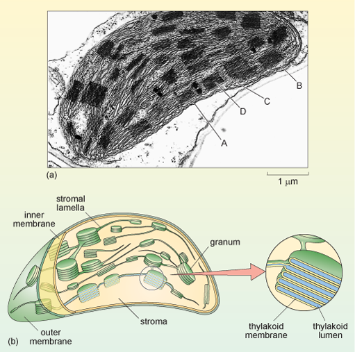
Electron micrograph of isolated chloroplasts with the major ...
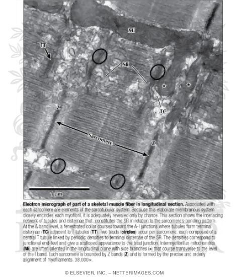
Electron Micrograph of Part of A Skeletal Muscle Fiber In ...

Biology: 1.2 Ultrastructure of Cells Flashcards | Quizlet

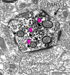
A-D Electron micrograph of HRP-labeled terminals in the ...
A tour of the cell: View as single page
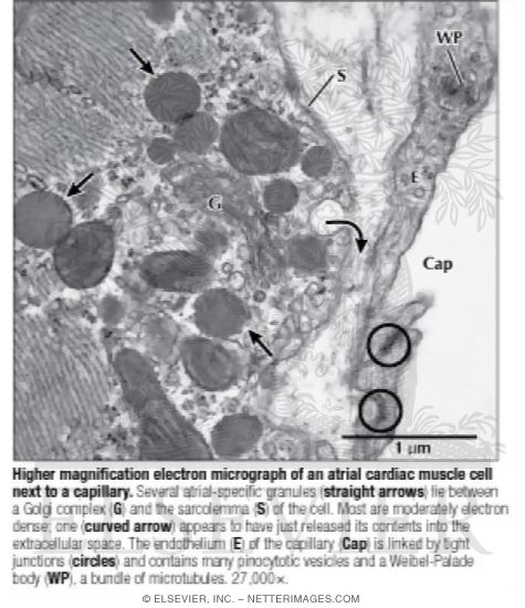
Higher Magnification Electron Micrograph of an Atrial Cardiac ...
Neuroanatomy Electron Microscopy Core | Feil Family Brain ...
DP Topic 1.1 / 1.2 | Biology - Quizizz
A tour of the cell: View as single page

USMLE Pathology Slides — Cell structures, electron microscopy ...
What is a diagram of a plant and animal cell under an ...
Post a Comment for "41 electron micrograph labeled"