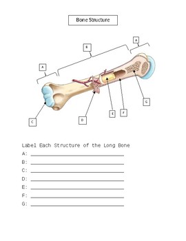
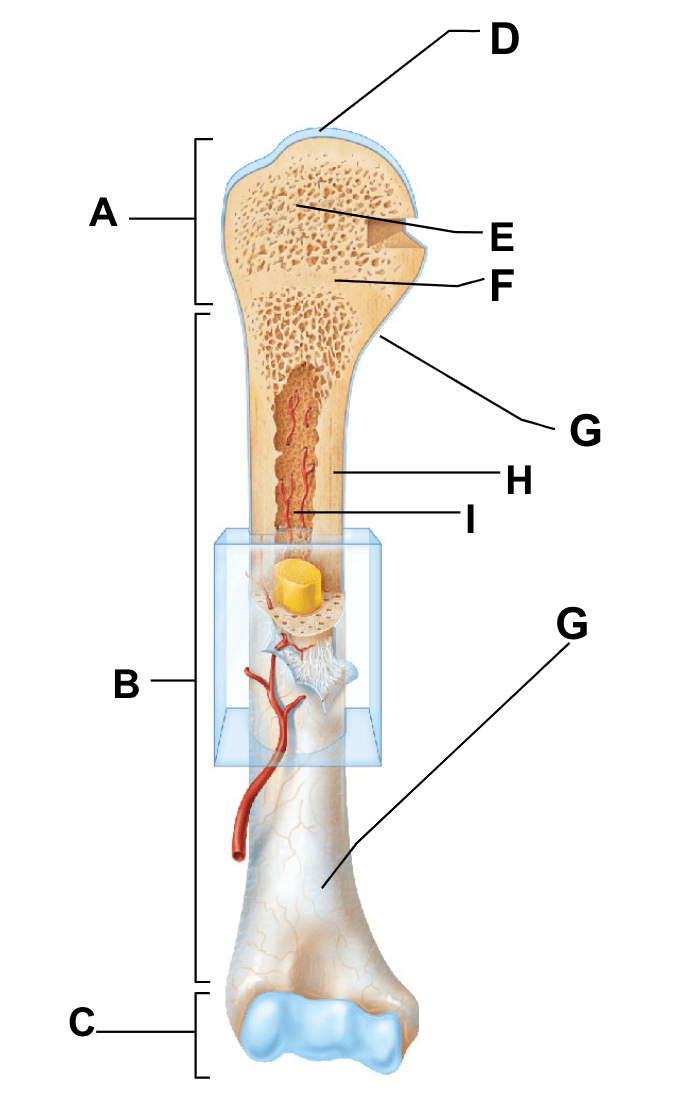
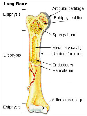
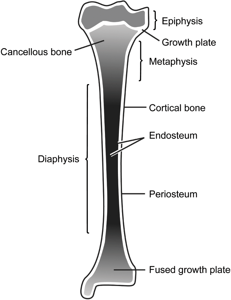
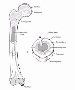
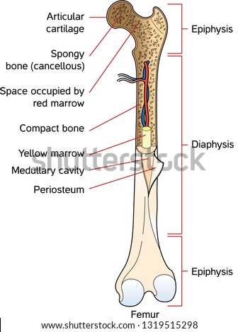
39 label the parts of the long bone
Learn femur anatomy fast with these femur quizzes | Kenhub Basic anatomy and parts of the femur The femur is a long bone found in the lower extremity. It serves as the attachment site for several muscles of the hip and leg, allowing it to withhold pressure from multiple angles. There are three main parts to the femur: The proximal end The shaft The distal end Scapula - Parts, Anatomy, Location, Functions, & Labeled Diagram The scapula, alternatively known as the shoulder blade, is a thin, flat, roughly triangular-shaped bone placed on either side of the upper back. This bone, along with the clavicle and the manubrium of the sternum, composes the pectoral (shoulder) girdle, connecting the upper limb of the appendicular skeleton to the axial skeleton.
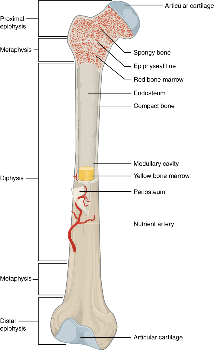
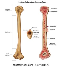
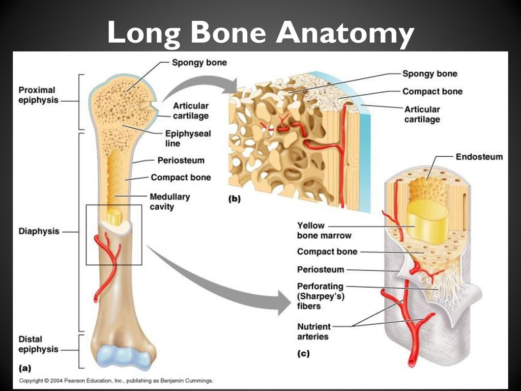
Bone markings [the complete list] | Kenhub Long bones are composed of four distinct parts: a head (epiphysis), a neck (metaphysis), a body (diaphysis), and an articular surface. The head, or epiphysis (epi- meaning "upon") of a bone refers to the rounded portion found at either ends of the bone.

Label the parts of the long bone
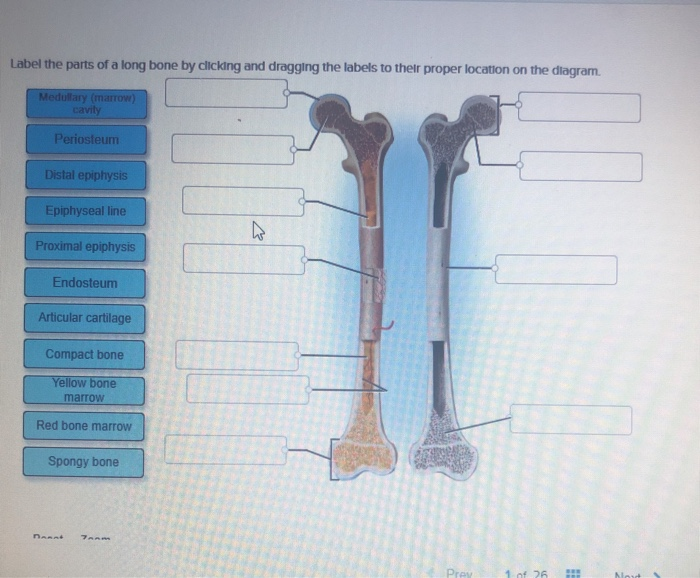
The Arches of the Foot - Longitudinal - Transverse - TeachMeAnatomy The foot has three arches: two longitudinal (medial and lateral) arches and one anterior transverse arch (Fig. 1).They are formed by the tarsal and metatarsal bones, and supported by ligaments and tendons in the foot. Their shape allows them to act in the same way as a spring, bearing the weight of the body and absorbing the shock produced during locomotion. The Four Types of Bone - Verywell Health Long bones of the leg include the femur, tibia, fibula, metatarsals, and phalanges. The clavicles (collar bones) are also long bones. Long bones provide the leverage we need for moving our bodies and for manipulating our environment. All long bones have two main parts: diaphysis and epiphysis. Diaphysis (Solved) - Label the parts of a long bone by clicking and dragging the ... Label the parts of a long bone ...
Label the parts of the long bone. What is Bone cancer? - All your info about health and medicine Cancer is a disease in which cells rapidly grow and divide to form a lump or mass. Cancer cells can spread to other parts of the body. Bone cancer is cancer that starts in bone cells. It may also start in cells of other bones. Cancer is a common cause of death in the United States. The risk of developing bone cancer increases with age. Horse Skeleton Anatomy - Osteological Features of Bones from Equine ... The humerus is a long bone of horse skeleton anatomy that possess some peculiar osteological features. It consists of a body and two different extremities. The body or shaft is irregularly cylindrical and has a twisted appearance. There presence four different surfaces in the shaft of the humerus of a horse. Wrist Bones: Anatomy, Function, and Injuries - Healthline Carpal bones in the wrist. Your wrist is made up of eight small bones called the carpal bones, or the carpus. These irregularly shaped bones join your hand to the two long forearm bones: the ... AccelStim Bone Growth Stimulator - P210035 | FDA The AccelStim Bone Growth Stimulator is intended to treat bone fractures that are not healing on their own. Specifically, it is used to help bone fractures of the larger bone between the elbow and ...
Bone Markings Quiz Questions And Answers - ProProfs Quiz Create your own Quiz. Here is this Bone markings quiz. In the study of anatomy from a functional and evolutionary standpoint, bone markings are used to identify individual bones and bony pieces. They are used by various professionals such as detectives, forensic scientists, osteologists, anatomists, radiologists, surgeons, and clinicians. Leg Bones Anatomy, Names & Diagram | Leg & Foot Bones - Study.com It can be defined either as the part between the hip bone and the ankle or as the part from the hip bone all the way down to the foot. In this lesson, the leg will comprise the entire length... The Osteon Or Haversian System Quiz Questions - ProProfs Quiz What is the strongest and longest bone in the body? A. Mandible B. Femur C. Humerus D. Tibia E. Ulna 2. What is the difference between endochondral and intramembranous ossification? A. In endochondral ossification, cartilage is replaced with bone, and in intramembranous ossification, mesenchyme in an embryo is transformed into bone. B. Axial Skeleton Anatomy: Diagram, Definition, Functions - Embibe Zygomatic bone: This pair of bones form the cheek of the face that articulates with sphenoid, temporal, and maxilla bones. Lacrimal bone: These pairs of bones form the part of the medial wall of the orbit. They are the smallest bone on the face. Nasal bone: These bones are two slender bones located at the front of the nose.
Labeled imaging anatomy cases | Radiology Reference Article ... X-ray cervical spine: lateral. X-ray cervical spine: AP. X-ray cervical spine: open-mouth peg. X-ray thoracic spine: frontal and lateral. X-ray lumbar spine: oblique. X-ray sacrum: frontal. CT cervical spine: bone window axial. CT cervical spine: bone window sagittal. CT cervical spine: bone window coronal. Anatomy, Bone Markings - StatPearls - NCBI Bookshelf Diaphysis - Refers to the main part of the shaft of a long bone. Long bones, including the femur, humerus, and tibia, all have a shaft. ... Santosh KC, Hegadi RS. Automated Fractured Bone Segmentation and Labeling from CT Images. J Med Syst. 2019 Feb 02; 43 (3):60. [PubMed: 30710217] 4. Plotkin LI, Essex AL, Davis HM. RAGE Signaling in Skeletal ... 206 Bone List PDF - Human All Bones Name with Picture - AIEMD Here is the complete list of all human bones. Axial Skeleton (80) Skull (28) Paired Bones (11 x 2 = 22) Nasal Lacrimal Inferior Nasal Concha Maxiallary Zygomatic Temporal Palatine Parietal Malleus Incus Stapes Frontal Ethmoid Vomer Sphenoid Mandible Occipital Torso (52) Paired Bones (12 x 2 = 24) Rib 1 Rib 2 Rib 3 Rib 4 Rib 5 Rib 6 Rib 7 The Ilium: Anatomy, Function, and Treatment - Verywell Health Anatomically speaking, the ilium is broken down into two parts: the body and the wing. The body of the ilium is its more central portion, and it forms a part of the acetabulum—the socket joint where the head of the femur (upper leg bone) rests—as well as the acetabular fossa, a deeper depression just above the joint. 1
Sketch And Label Of A Cross Section Of A Long Bone In long bones, as you move from the outer cortical compact bone to the inner medullary cavity, the bone transitions to spongy bone. Lamellar bone makes up the compact or cortical bone in the skeleton, such as the long bones of the legs and arms. Label lines should not cross. Sketch and label a cross section of a bone.
A Picture Guide to the Different Parts of a Horse Scroll through the photographs for a closer look at each body part. Identified for you are the: Muzzle Poll Forelock Ears Eyes Forehead Nostrils Cheek Neck Shoulder Forearm Knee Front Cannon Bone Fetlock Pastern Back Barrel Loins Flanks Gaskin Stifle Hock Hind Cannon Bone Croup Dock Tail 01 of 29 Muzzle Friederike Von Gilsa/Getty Images
Skeletal System: Types of Bones in the Human Body - EZmed First we said to remember "long" and "limb" because most of the long bones can be found in the arms and legs including: Upper Extremity Humerus (2) - Arm Radius (2) - Forearm Ulna (2) - Forearm Metacarpals (10) - Hand Phalanges (28) Fingers Lower Extremity Femur (2) - Upper Leg Tibia (2) - Lower Leg Fibula (2) - Lower Leg Metatarsals (10) - Foot
Chicken Anatomy 101: Everything You Need To Know The skull, humerus (arm bone), pelvis, and collar bones are examples Medullary: These bones store calcium. In the centers of these bones is bone marrow which makes blood cells. Legs, shoulder blades, and ribs are examples of this type. The neck and backbone of the chicken are very flexible.
Chicken Skeleton Anatomy with Labeled Diagram In the labeled diagram, I showed you the skull bone, vertebrae (cervical to caudal), ribs, sternum, wing bones, and leg bones from a chicken. You may also get help from the video that I will add at the end of this article. That video might help you to identify all the bones of a chicken. Chicken skeleton anatomy diagram Chicken bone anatomy
Clavicle - Definition, Location, Anatomy, & Labeled Diagram Anatomy - Parts of the Clavicle Along With its Bony Landmarks Being a long bone, clavicle has two ends, sternal and acromial end. The region in between the two ends is known as shaft. C l a v i c l e 1. Sternal (Medial) End The part of the clavicle that lies towards the sternum is called the sternal end or the medial end.
Major Landmarks of Bones - Video & Lesson Transcript | Study.com The enlarged end of a long bone is called the epiphysis. The end of a bone may have other features on it, but the whole thing is called an epiphysis. Always remember that epiphysis is an enlarged...
What Do Dogs and Other Mammals Have That Humans Don't? A Penis Bone Penis bone of the Japanese macaque. (Didier Descouens / CC BY-SA 4.0 ) The label is descriptive. A penis bone is a bone in the penis. It belongs to a classification of heterotopic bones or bones that are dissociated from the skeleton. In most cases, it rests in the abdomen until sexual arousal causes it to extend within the erectile tissue of ...
(Solved) - Label the parts of a typical long bone on the following ... Label the parts of a typical ...
Humerus Bone Anatomy, Function, Fractures, More - Healthline as a long bone. Other types of long bones include the radius and ulna in your forearm and the femur in your upper leg. Speaking of long, the humerus is the longest bone in your arm. Despite its...
Vagina Parts | a Diagram and Guide of Female Anatomy 1. The vulva. It's a common misconception that the visible outer parts of the female anatomy is called the vagina. The technical name is actually the vulva. Yours has the job of protecting your ...
(Solved) - Label the parts of a long bone by clicking and dragging the ... Label the parts of a long bone ...
The Four Types of Bone - Verywell Health Long bones of the leg include the femur, tibia, fibula, metatarsals, and phalanges. The clavicles (collar bones) are also long bones. Long bones provide the leverage we need for moving our bodies and for manipulating our environment. All long bones have two main parts: diaphysis and epiphysis. Diaphysis
The Arches of the Foot - Longitudinal - Transverse - TeachMeAnatomy The foot has three arches: two longitudinal (medial and lateral) arches and one anterior transverse arch (Fig. 1).They are formed by the tarsal and metatarsal bones, and supported by ligaments and tendons in the foot. Their shape allows them to act in the same way as a spring, bearing the weight of the body and absorbing the shock produced during locomotion.











Post a Comment for "39 label the parts of the long bone"