42 skull diagram labeled
Learn skull anatomy with skull bone quizzes and diagrams 28/10/2021 · Skull anatomy diagrams. As mentioned, the skull is home to so many structures that the prospect of learning them all can seem very overwhelming. An easy step-by-step system for breaking the topic down then, is essential. For this, we … What are the dermatomes of the head and neck? - Medscape Dermatomes of the Head and Neck (Open Table in a new window) Divisions of the trigeminal nerve (cranial nerve [CN] V1, V2, and V3) Most of the skin of the face, including anterior aspect of lower ...
Cat Skeleton Anatomy with Labeled Diagram - AnatomyLearner 29/05/2021 · Cat skull has a short fascial and palatal region compares to other mammals. The skull is oval elongated in shape, and has strong, highly curved zygomatic bones. There is an incomplete orbital rim in cat skull anatomy. Premaxilla is a separate bone in cat skull anatomy. Masseteric fossa present in the mandible of a cat (also found in a dog).
Skull diagram labeled
Functions of Human Skeletal System | Just-Health.net Functions of the Skeleton. The important functions make the human body superior to other living species. The human skeleton has special features, such as having opposable thumbs and walking erect on two legs, which make man unique and highly functional. To appreciate these, let us take a look at the vital functions of the skeleton: Shape. Organs of Skeletal System and Their Functions - New Health Advisor Skull- it includes the cranium, face and auditory ossicles. Hyoid - bone of neck for muscles attachment of chin and larynx. Vertebral column - consist of all spinal vertebrae. Thoracic cage - it contains ribs and sternum. Appendicular Skeleton It contains the following from top to bottom respectively: How to Work Safely with - Hazardous Products using the "Skull and ... What does this pictogram mean? The symbol within the pictogram is a human skull with two crossed bones behind it. The symbol indicates that hazardous products with this pictogram can cause death or poisoning. Hazardous products with this pictogram can be safely worked with if proper storage and handling practices are followed.
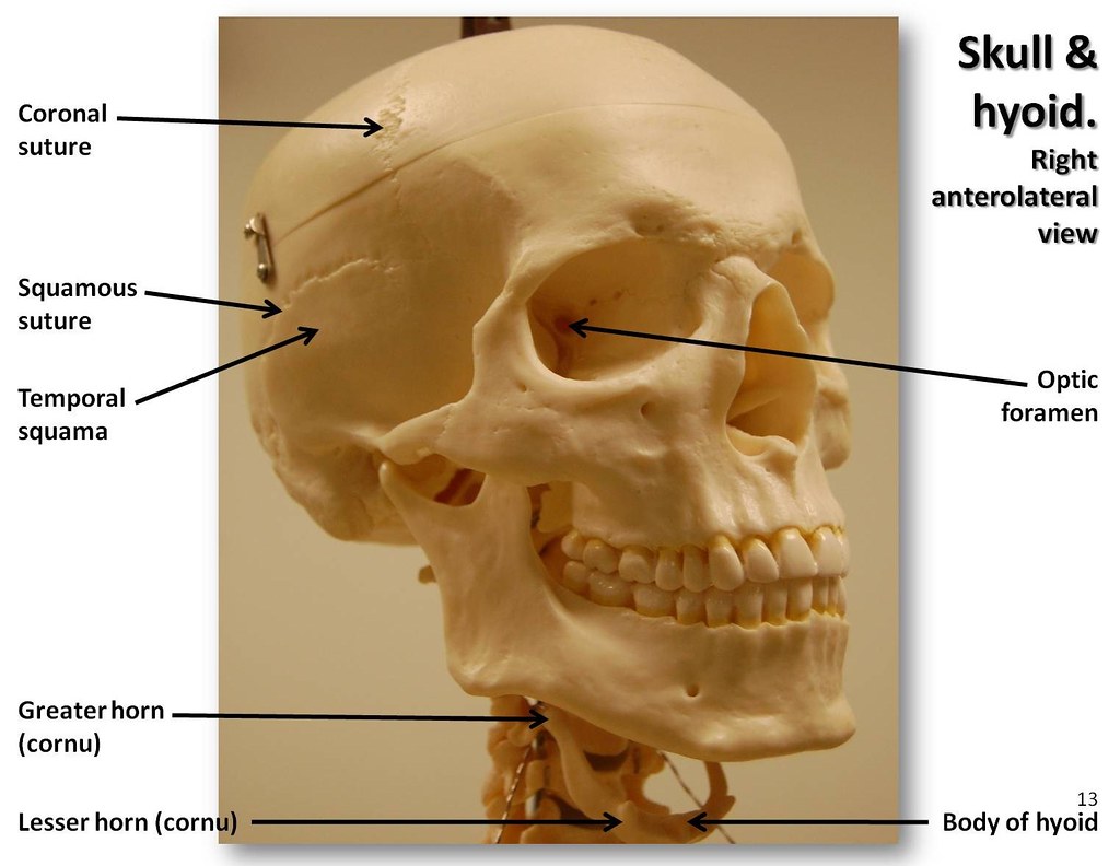
Skull diagram labeled. Sutures of the skull: Anatomy | Kenhub The sutures of the skull, also referred to as the cranial sutures, are fibrous joints that connect the bones of the skull. They appear as intricate thin lines that mark the adherence between the bones and the growth and closure of the cranial fontanelles. The dense fibrous tissue that connects the sutures is made mostly out of collagen. blank anatomy diagrams pdf blank anatomy diagrams pdf. wecc balancing authority map Posted on June 9, 2022 odessa, mo high school basketball By lawrence university the rock on blank anatomy diagrams pdf ... Positions and Functions of the Four Brain Lobes - MD-Health.com The brain is divided into four sections, known as lobes (as shown in the image). The frontal lobe, occipital lobe, parietal lobe, and temporal lobe have different locations and functions that support the responses and actions of the human body. Let's start by identifying where each lobe is positioned in the brain. Position of the Lobes Anatomy Project - Sheridan College Neck. · Connecting the shaft and head of the femur. · Projects superior and medial from the shaft to the head. · In addition to projecting superior and medial from the shaft of the femur, the neck also projects somewhat anterior. · The amount of forward projection is extremely variable, but on an average is from 12° to 14°.
Stem: Definition, Types, Functions and Diagram- Embibe Ans: 1. The primary functions of the stem are to provide support to various parts of the plant and transportation of water and minerals to leaves, flowers, buds, fruits, etc. 2. It promotes important processes such as photosynthesis, pollination, fertilization by holding leaves and plants in the right positions. 3. Dermoid Cyst: Practice Essentials, Epidemiology, Prognosis For pediatricians and dermatologists, dermoid cyst means subcutaneous cysts, which are usually congenital. [ 1] In all disciplines, however, the common factor is the presence of a solitary, or occasionally multiple, hamartomatous tumor. The tumor is covered by a thick dermislike wall that contains multiple sebaceous glands and almost all skin ... anatomylearner.com › cat-skeleton-anatomyCat Skeleton Anatomy with Labeled Diagram May 29, 2021 · Cat skull has a short fascial and palatal region compares to other mammals. The skull is oval elongated in shape, and has strong, highly curved zygomatic bones. There is an incomplete orbital rim in cat skull anatomy. Premaxilla is a separate bone in cat skull anatomy. Masseteric fossa present in the mandible of a cat (also found in a dog). Organs of the body | Their Locations and Internal Functions This is the master organ of the body. All the organ systems of the human body are under its control. The skull, a bone frame in the head, houses the brain. It is made up of nerve cells and neuroglia. It consists of parts like the cortex, cerebral hemisphere, cerebellum, medulla oblongata, and pons. It extends into the spinal cord.
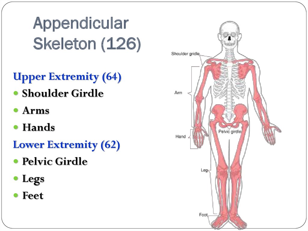
› blog › skull-facial-bonesFacial Bones of the Skull Mnemonic: Anatomy and Labeled ... Nov 17, 2020 · Use this facial bone mnemonic list to remember the anatomy, names, and structure of each of the facial bones of the skull. Uses labeled diagrams to show the structure and anatomy of each facial bone. Includes discussion about features and functions of the maxillary, mandible, nasal, conchae, zygomatic, vomer, lacrimal, and palatine bones. › pin › 182818066098974005Printable Human Skeleton Diagram - Labeled, Unlabeled, and ... Oct 25, 2014 - Click here to download a free human skeleton diagram. Great for artists and students studying human anatomy. Includes labeled human skeleton chart. Appendicular Skeleton: Bones List, Diagram & More - Embibe Figure: The Upper Limbs. 4.The Lower limbs or hind arm: The lower limb contains a total of \(60\) bones, i.e., \(30\) on each side.The lower limb consists of the following bones: femur, tibia, fibula, patella, tarsals, metatarsals, and phalanges. (1) Femur: It is the longest bone of the body and the only bone in the thigh. It is present in the lower limb between the hip and the knee joint. Rare-earth based materials: an effective toolbox for brain imaging ... A high performance Sc-based nanoprobe for through-skull fluorescence imaging of brain vessels beyond 1500 nm. Nanoscale 10 , 9393-9400 (2018). Google Scholar
Skull, anterolateral view with labels - Axial Skeleton Vis… | Flickr
Diatom - Wikipedia Diatoms are classified as eukaryotes, organisms with a membrane-bound cell nucleus, that separates them from the prokaryotes archaea and bacteria. Diatoms are a type of plankton called phytoplankton, the most common of the plankton types. Diatoms also grow attached to benthic substrates, floating debris, and on macrophytes.

Skull Medial view of sagittal region Quiz
Parts and Components of Human Ear and Their Functions The outer ear is the portion of the ear that sits atop the skull, which is made of flesh and cartilage. It is the visible part which serves to protect the eardrum. It also collects and guides sound waves into the middle ear. Compositional parts and their functions . Pinna (ear flap) The ear flap or pinna is the outer portion of the ear.

121462.jpg
Dog Skeleton Anatomy with Labeled Diagram 31/12/2021 · Here, in the dog skeleton labeled diagram, I tried to show you the different segments of the forelimb, hindlimb with their bones. Again, I tried to show you all the bones from the vertebrae column of a dog skeleton. In addition, in the diagram, you will find a few identified skull bones. The sternum and the ribs are also identified in the dog ...
The Skeletal System - презентация онлайн
What are the 12 cranial nerves? Functions and diagram Each nerve has a name that reflects its function and a number according to its location in the brain. Scientists use Roman numerals from I to XII to label the cranial nerves in the brain. The 12...

Inside of skull labeled | Diş anatomisi, Nörobilim, Tıbbi
Skeletal muscle tissue: Histology - Kenhub Skeletal muscle is an excitable, contractile tissue responsible for maintaining posture and moving the orbits, together with the appendicular and axial skeletons. It attaches to bones and the orbits through tendons. Excitable tissue responds to stimuli through electrical signals. Contractile tissue is able to generate tension of force.
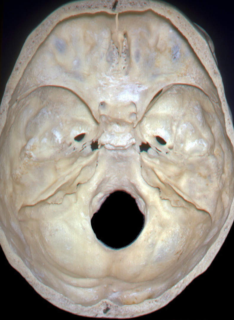
Internal Skull Base Anatomy | Neuroanatomy | The Neurosurgical Atlas
anatomylearner.com › dog-skeleton-anatomyDog Skeleton Anatomy with Labeled Diagram Dec 31, 2021 · In addition, in the diagram, you will find a few identified skull bones. The sternum and the ribs are also identified in the dog skeleton labeled diagram. If you want to more updated dog skeleton labeled diagram, you may join anatomy learner on social media (get more images). Frequently asked questions on dog skeleton bones. So, again this part ...
A
› en › libraryLearn skull anatomy with skull bone quizzes and diagrams - Kenhub Oct 28, 2021 · Skull anatomy diagrams. As mentioned, the skull is home to so many structures that the prospect of learning them all can seem very overwhelming. An easy step-by-step system for breaking the topic down then, is essential. For this, we love labeled diagrams. Labeled Skull Diagram
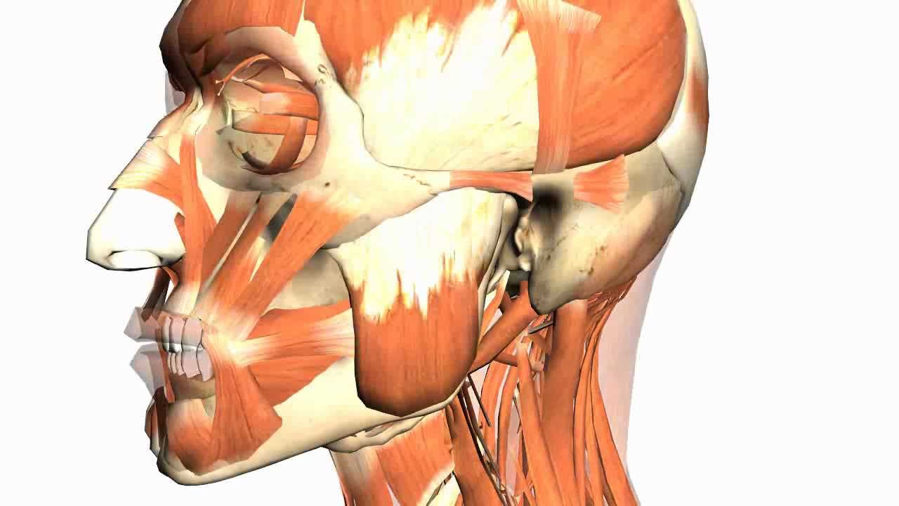
Skull tutorial (4) - Mandible - Anatomy Tutorial - YouTube
A List of Bones in the Human Body With Labeled Diagrams The skull is supported by the first cervical vertebra, which is known as the atlas. The second cervical vertebra, known as the axis, forms the pivot on which the atlas turns. The cervical vertebrae form the neck. The cervical vertebrae are designated as C1 to C7, as shown in the diagram. Thoracic Vertebrae

Anterior muscle anatomy | Muscular system, Muscular system labeled ...
Antenatal Care Module: 6. Anatomy of the Female Pelvis and Fetal Skull ... The fetal skull bones are as follows: The frontal bone, which forms the forehead. In the fetus, the frontal bone is in two halves, which fuse (join) into a single bone after the age of eight years. The two parietal bones, which lie on either side of the skull and occupy most of the skull. Parietal is pronounced 'parr eye ett al'.
Post a Comment for "42 skull diagram labeled"