43 label compound microscope
Label Parts Of A Compound Microscope Teaching Resources | TpT This is from a set of 3 tiered readings. Students will read a passage about the how to use a compound light microscope. Students will use textual evidence to answer questions and label the different parts of the microscope. It also allows students to gain prior knowledge about the compound microscope. Version A provides the most support for ... BYJUS BYJUS
PDF Label compound microscope worksheet Label compound microscope worksheet Name_____. In paragraph 15, when you concentrating the sample, you should always start with the goal of _____ 16. Only the _____ button should be used when using a high-power target. 17. The type of microscope used in most science classes is the _____ microscope.

Label compound microscope
How To Make Slime: 4 Best Slime Recipes | Science Projects Label an 8 oz. plastic bottle “Sodium Tetra-borate (Borax) Solution” with a permanent marker. Fill the bottle about ¾ full with water. Add 4 teaspoons of sodium tetraborate to the water and shake until mostly dissolved. Fill the bottle to the top with water and shake again to completely dissolve the sodium tetraborate solids. Cardiomyocytes (Cardiac Muscle Cells)- Structure, Function In order to observe cardiomyocytes under the microscope, it is necessary to fix and attach the cells on a microscope. Once the cells have been fixed and permeabilized on the slide, then they are ready for staining and viewing. Requirements A sample of cardiomyocytes (obtained from such an animal as rodents) Paraformaldehyde › article › how-to-make-slimeHow To Make Slime: 4 Best Slime Recipes | Science Projects Label an 8 oz. plastic bottle “Sodium Tetra-borate (Borax) Solution” with a permanent marker. Fill the bottle about ¾ full with water. Add 4 teaspoons of sodium tetraborate to the water and shake until mostly dissolved. Fill the bottle to the top with water and shake again to completely dissolve the sodium tetraborate solids.
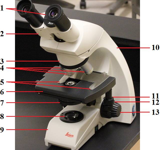
Label compound microscope. Parts of a Compound Microscope (And their Functions) List of Microscope Parts and their Functions. 1. Ocular Tubes (Monocular, Binocular & Trinocular) The ocular tubes, are to tubes that lead from the head of the microscope out to your eyes. On the end of the ocular tubes are usually interchangeable eyepieces (commonly 10X and 20X) that increase magnification. Label the Parts of a Compound Light microscope - BIOLOGY JUNCTION Label the Parts of a Compound Light microscope Parts of the Compound Microscope - HCC Learning Web Parts of the Compound Microscope Use Figure 2 as a guide to locate the major parts of the compound microscope. a. Base: The bottom, flat part that supports the microscope. b. Arm: The straight or curved vertical part that connects the base to the upper portion. c. Body Tube: Extends from the arm and contains the ocular lens and the rotating Parts of a microscope with functions and labeled diagram Head - This is also known as the body. It carries the optical parts in the upper part of the microscope. Base - It acts as microscopes support. It also carries microscopic illuminators. Arms - This is the part connecting the base and to the head and the eyepiece tube to the base of the microscope.
Solved Label the image of a compound light microscope using - Chegg Experts are tested by Chegg as specialists in their subject area. We review their content and use your feedback to keep the quality high. Transcribed image text: Label the image of a compound light microscope using the terms provided. p-Cresol | CH3C6H4OH - PubChem compound Summary; p-Cresol. Cite. Download. PubChem CID: 2879: Structure: Find Similar Structures. Chemical Safety: Laboratory Chemical Safety Summary (LCSS) Datasheet. Molecular Formula: C 7 H 8 O or CH 3 C 6 H 4 OH: Synonyms: P-CRESOL. 4-Methylphenol. 106-44-5. 4-Cresol. 4-Hydroxytoluene. More... Molecular Weight: 108.14. Dates: Modify . 2022-05-20 . … Compound Microscope Labeled Diagram | Quizlet QUESTION. The total magnification of a specimen being viewed with a 10X ocular lens and a 40X objective lens is. 15 answers. QUESTION. a mosquito beats its wings up and down 600 times per second, which you hear as a very annoying 600 Hz sound. if the air outside is 20 C, how far would a sound wave travel between wing beats. 2 answers. researchtweet.com › microscope-parts-labeledMicroscope, Microscope Parts, Labeled Diagram, and Functions Jan 19, 2022 · Simply multiply the magnification of the ocular lens by the magnification of the objective lens to calculate the power of magnification of a microscope. For a typical compound microscope with a 10X ocular lens and objective lenses with magnifications of 4X, 10X, 40X, and 100X, your microscope will have magnifications of 40X, 100X, 400X, and ...
Parts of the Microscope with Labeling (also Free Printouts) Parts of the Microscope with Labeling (also Free Printouts) A microscope is one of the invaluable tools in the laboratory setting. It is used to observe things that cannot be seen by the naked eye. Table of Contents 1. Eyepiece 2. Body tube/Head 3. Turret/Nose piece 4. Objective lenses 5. Knobs (fine and coarse) 6. Stage and stage clips 7. Aperture pubchem.ncbi.nlm.nih.gov › compound › p-cresolp-Cresol | CH3C6H4OH - PubChem P-cresol is a cresol that consists of toluene substituted by a hydroxy group at position 4. It is a metabolite of aromatic amino acid metabolism produced by intestinal microflora in humans and animals. Compound Microscope- Definition, Labeled Diagram, Principle, Parts, Uses A compound microscope is of great use in pathology labs so as to identify diseases. Various crime cases are detected and solved by drawing out human cells and examining them under the microscope in forensic laboratories. The presence or absence of minerals and the presence of metals can be identified using compound microscopes. Compound Microscope - Diagram (Parts labelled), Principle and Uses Compound Microscope - Diagram (Parts labelled), Principle and Uses Compound Microscope As the name suggests, a compound microscope uses a combination of lenses coupled with an artificial light source to magnify an object at various zoom levels to study the object. A compound microscope: Is used to view samples that are not visible to the naked eye

Chapter 7, Page 8 - HistologyOLM
10 Best Compound Microscopes (Spring 2022) - The Complete Guide Celestron Labs designed a premium compound microscope with exceptional durability. It comes with a two-year warranty, but it will last longer if you maintain it right. The product includes two eyepieces, a WP-20x and a WF-10x with a pointer. You can change these eyepieces easily to find your perfect adjustment.
leica_dm500.jpg
› panoramic › indexPharmaCircle This website uses cookies to help provide you with the best possible online experience. Please read our Terms & Conditions and Privacy Policy for information about ...
Life cycle Laminaria and Fucus (Brown Algae)
Parts of a Compound Microscope and Their Functions Mechanical Parts. Compound microscope mechanical parts include base or foot, pillar, arm, inclination joint, stage, clips, diaphragm, body tube, nose piece, coarse adjustment knob and fine adjustment knob. Base: It's the horseshoe-shaped base structure.
Light Microscopy and Photomicrography
What are the parts of a microscope labeled? - SidmartinBio (A) Mechanical Parts of a Compound Microscope Foot or base. It is a U-shaped structure and supports the entire weight of the compound microscope. Pillar. It is a vertical projection. Arm. The entire microscope is handled by a strong and curved structure known as the arm. Stage. Inclination joint. Clips. Diaphragm. Nose piece.
Unit 2: Microscopes - Biology 1C/2C 2012-13
Compound Microscope Parts, Functions, and Labeled Diagram The total magnification of a compound microscope is calculated by multiplying the objective lens magnification by the eyepiece magnification level. So, a compound microscope with a 10x eyepiece magnification looking through the 40x objective lens has a total magnification of 400x (10 x 40).

Chapter 6, Page 5 - HistologyOLM
Label the microscope — Science Learning Hub All microscopes share features in common. In this interactive, you can label the different parts of a microscope. Use this with the Microscope parts activity to help students identify and label the main parts of a microscope and then describe their functions. Drag and drop the text labels onto the microscope diagram.

Compound Light Microscope Labeled - Made By Creative Label
compound microscope parts (labeling) Flashcards | Quizlet Only $35.99/year compound microscope parts (labeling) STUDY Flashcards Learn Write Spell Test PLAY Match Gravity Created by barnettlily Terms in this set (14) eyepiece tube - connects the eyepiece to the objective lens what is 1? nosepiece (turret) - holds and spins the objective lenses what is 2?
Personal Experience with Microscopes - AyushiSinhaMicroscopy
Quia - Label the Microscope Quiz Label the Microscope Quiz. Choose the word that correctly labels the parts of the microscope. Please enter your name. First name: Last name . Tools. Copy this to my account; E-mail to a friend; Find other activities; Start over; Print; Help; Mrs. Coyle. Simmons Elementary School. Versailles, KY: View profile;

Microscope Parts
Cole-Parmer China, an Antylia Scientific company - Lab equipment … Cole-Parmer, an Antylia Scientific company, provides the widest range of lab equipment and supplies for pharmaceutical, biopharma, healthcare, and environmental.

Labeling a Compound Microscope - PurposeGames
Microscope Parts and Functions First, the purpose of a microscope is to magnify a small object or to magnify the fine details of a larger object in order to examine minute specimens that cannot be seen by the naked eye. Here are the important compound microscope parts... Eyepiece: The lens the viewer looks through to see the specimen.

OMAX Microscope 40X-1000X Student Compound Microscope with Tungsten Light
Labeling the Parts of the Microscope Labeling the Parts of the Microscope This activity has been designed for use in homes and schools. Each microscope layout (both blank and the version with answers) are available as PDF downloads. You can view a more in-depth review of each part of the microscope here. Download the Label the Parts of the Microscope PDF printable version here.
Post a Comment for "43 label compound microscope"